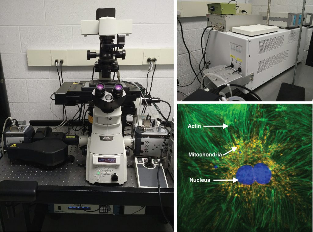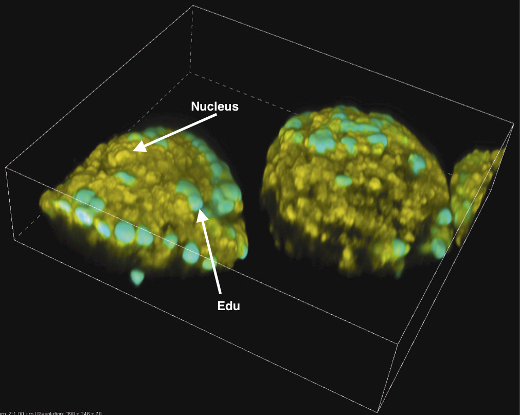
The 3D Facility SFC Microscope enables dynamic real time live cell imaging with a high-sensitivity & high-speed confocal fluorescence microscope in a full Biosafety level 2 room. The slit scanning mode of the SFC microscope produces faster acquisition and lower laser driven toxicity.
Instrument details:
• Powerful laser lines: 405nm, 488nm, 561nm, 640nm
• Motorized components for automated acquisition
• Live cell chamber: temperature & CO2 control environment
• Easy to use: z-stack, multipoint, large image XY stitching applications for ultra-high definition, time-lapse imaging automated cell counting, fluorescence intensity analysis, morphometric measurements
• Real time confocal imaging at speed up to 30 fps with the SFC module
• Nikon perfect focus to minimize focus drift during time lapse acquisition
• Full integration with NIS Elements software makes the SFC module very easy to learn and use
Applications:
• 3D real time imaging
• volume visualization & analysis studies
• Live cell imaging
• Well plate scanning
• Cell counting
• Cell viability, proliferation, autophagy, cytotoxicity, metabolism
• Cell structure
• Calcium transient studies
• Fast neural events
• Colocalization studies
• Cell surface event tracking, localization & colocalization
And many more….

