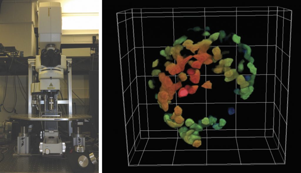
The 3D Facility Nikon A1R 2-photon microscope is an ultra-fast, multi-color instrument designed for a multitude of in-vivo imaging applications in living specimens in a full Biosafety level 2 room.
Instrument details:
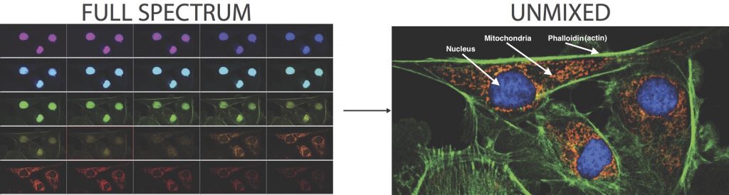
• Spectral detector in 2-photon mode: image up to 6 different fluorescent probes at once.
• IR non-descanned PMT detector: high-sensitivity & high-resolution
• Resonant scanner: ultrafast imaging up to 400 frames/sec
• High-power & tunable 700 – 1000nm Ti:Sapphire laser (with auto alignment feature) for deep imaging
• Stage Piezoelectric micrometer Z drive & Objective Nano positioning system: very fast and very accurate 3D imaging and positioning
• Fully customizable stage for in-vivo studies with ventilator assisted anesthesia capabilities
• Stereotaxic instrument available for in-vivo imaging on rodent head
Applications:
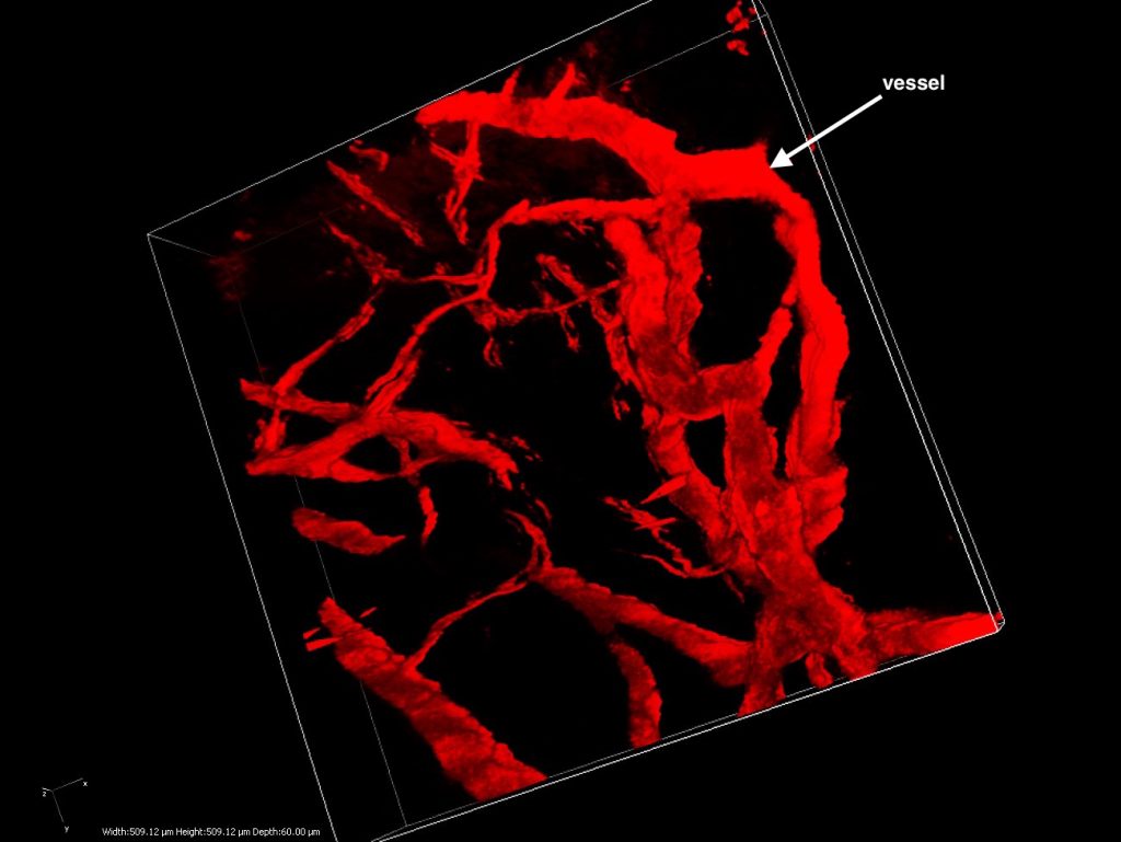
• 3D and volume imaging for visualization & analysis studies
• In-vivo imaging studies
• Live cell studies
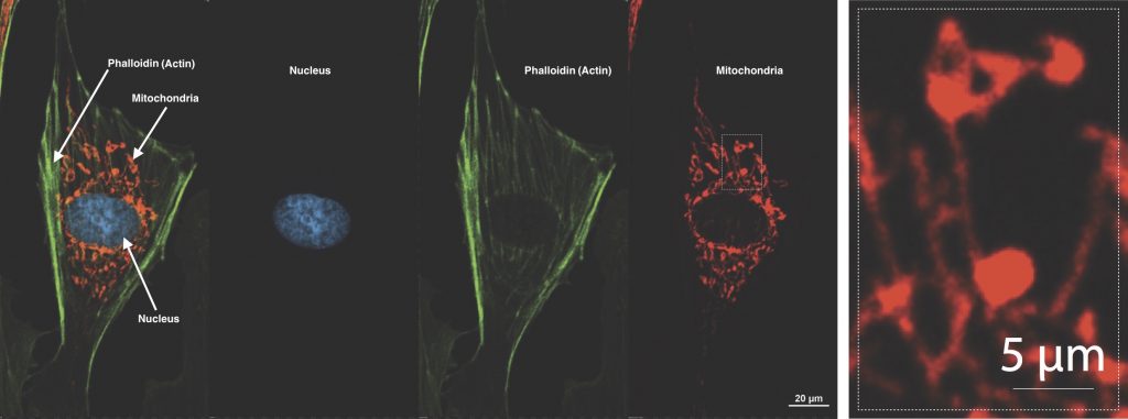
• Cell structure studies with high resolution pictures
• Ultrafast calcium transient studies
• Voltage-sensitive fluorescent studies
• Colocalization studies
And many more….