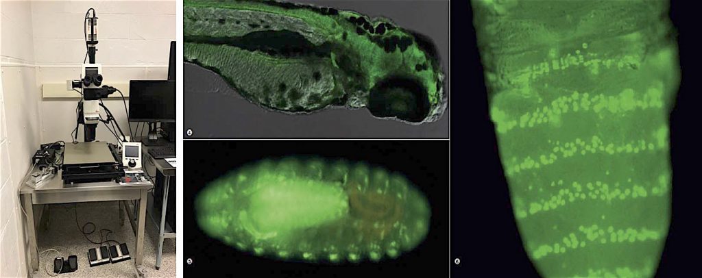
promoter. Source – Leica-Prof. Dr. Stephan C. F. Neuhauss, University of Zurich; middle bottom: Peripheric and central nervous (ventral cord) system of a drosophila embryo, salivary gland; right: Drosophila melanogaster. Dorsal view, Pupa; Green: Venus. Transgenic
fluorescent protein in posterior compartment of each segment. Source – Leica-Dr. Kuranaga, The University of Tokyo
The Facility Leica M205 is the perfect instrument for procedures requiring long working distance such as surgeries. This stereomiscroscope housed in a full Biosafety level 2 room is also multimodal & fully automatized for observation, picture and video acquisition of procedures. It also includes a complete epifluorescent mode to multiplex with the brightfield. The optional 3D capability add-on is part of this setup which allows deconvolution, visualization and measurements in 3D of macroscale biological samples.
Instrument details:
• Magnification ranges from 0.5x-5x with long working distance of 139mm
• Powerful 100W mercury lamp illumination multicolor fluorescent experiment
• Versatile & automatized Fluorescence modality: separate illumination beam path for fluorescence, motorized four position fluorescence filter turret, motorized fluorescence illumination shutter, motorized fluorescence intensity control, motorized iris diaphragm
• Excitation filters: 350nm, 470nm, 560nm; Emission filters: 460nm, 525nm, 630nm
• Up to 5 acquisition channels can be define per experiment
• Hand free support with the fully software configurable Leica Footswitch (foot pedals control the zoom & focus)
• 2 Leica cameras for 2 different acquisition paths:
– a Leica MC190 HD (10 Megapixels) color camera to teach and record video of surgery (or other procedures)
– a deep-cooled Leica DFC9000 GT (4 Megapixels) to take high resolution pictures in brightfield or in fluorescent mode
• The LAS X core software provide full customization of the stereomicroscope controls, allows powerful acquisition, visualization and analysis of videos and pictures.
• The LAS X software module controls the Z focus motor, SuperZ finefocus, define focus positions to capture 3-dimensional data sets
• The software package LAS X 3D Surface add-on allows generation, reviewing and measurement of 3D surface views originating from image z stacks (extended depth of field, 3D surface viewer, 3D Surface measurement)
Applications:
• Teachable surgery techniques by acquisition of high definition pictures and movie sequences in live stream mode
• 2D & 3D acquisition for the study of macroscale biological samples
• 3D volume measurements of macroscale biological samples
Fluorescent imaging & analysis of macroscale biological samples
And many more…